Epidemiology of scoliosis, who is most at risk?
Scoliosis is the most common orthopedic condition among children and adolescents, with an estimated prevalence of up to 15.3%. Literature reports similar occurrence rates of frontal plane spinal deformities ranging from 6° to 10° in both boys and girls. However, when the curvature exceeds 21°, the ratio of girls to boys is 5.4 to 1. According to Głowacki et al., for every boy with a Cobb angle in the range of 20°-30°, there are up to 10 girls. The prevalence of scoliosis is dependent on age and gender.
Etiology of scoliosis, where does it come from?
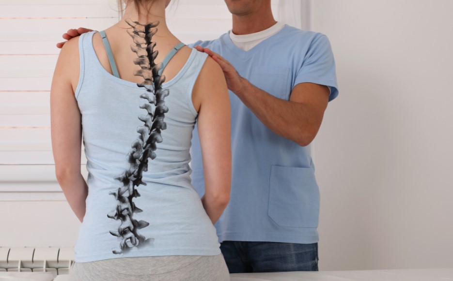
In some cases, the cause of scoliosis can be determined, but in many cases, the etiology remains unclear. It can be related to factors such as limb length discrepancy, pain, muscle weakness, or congenital abnormalities. The development of lateral spinal curvature may also be associated with an oblique pelvic tilt, which changes the distribution of forces in the hip joints and the lumbar spine. When the vertebrae are misaligned in the frontal plane, it disrupts the axis of rotation, which is in the transverse plane. The gravitational force acting on this abnormal axis generates rotational forces on the vertebrae. This rotation can lead to the development of lateral spinal curvature and its associated consequences.
Importantly, the largest group of scoliosis cases, accounting for up to 90%, is idiopathic (of unknown cause). There are only hypotheses regarding the factors that may contribute to their development. Genetic factors, early exposure to toxins, and hormonal imbalances are believed to play a role. Biomechanics, lifestyle factors, and environmental conditions are also considered as potential causes [6,7]. Researchers in this field have developed various theories to explain the etiology of scoliosis based on clinical observations and studies. However, none of these theories have been fully confirmed.
Classification of scoliosis
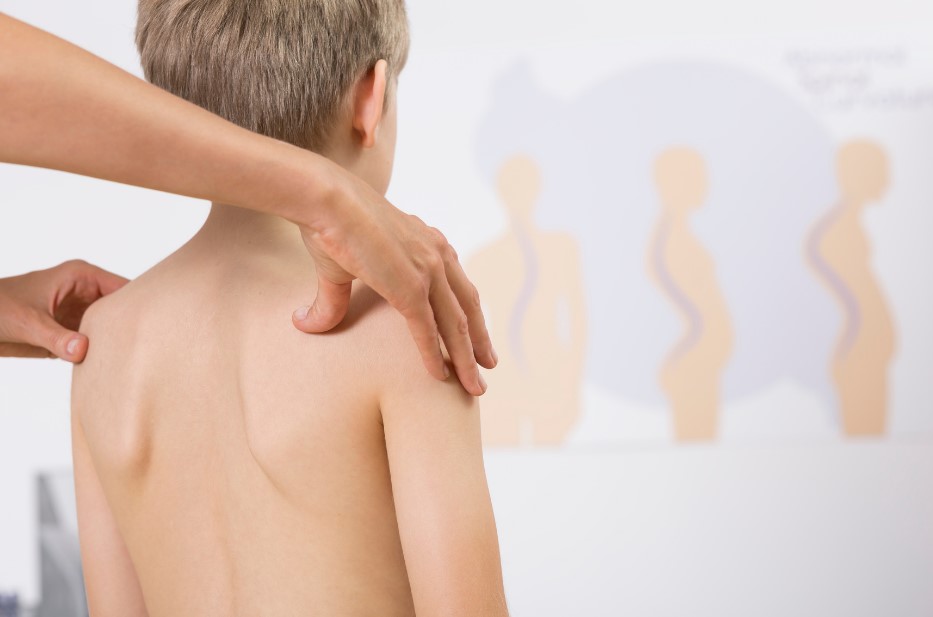
Scoliosis can be classified based on various criteria, including chronology, location of the curvature, angular magnitude, and etiology. Wejsflog proposed a classification of scoliotic symptoms into three groups based on the degree of deformity in the musculoskeletal system. In first-order scoliotic symptoms, there is torsion and wedging of the vertebrae, as well as three-dimensional spinal curvature. Second-order symptoms involve skeletal elements directly associated with the spine. These include posterior rib hump on the convex side and rib depression on the concave side. On the anterior surface of the chest, the situation is reversed, with depression on the convex side and a rib hump on the concave side. Second-order symptoms also include lateral displacement of the hip, inclination of the chest, and its displacement, typically towards the convex side. Third-order changes involve further regions of the musculoskeletal system. These include deepening of the waist triangle on the concave side. Asymmetrical positioning of the shoulders and scapulae is observed, with the scapula on the convex side protruding and rotating outward.
Taking into account the chronology of scoliosis development, James proposed a classification. According to him, there are early-onset scoliosis, which occur around the age of 3, during the first growth spurt. These scoliosis types are more common in boys, and the deformity affects the thoracic spine. This type of scoliosis can be progressive or non-progressive. In the progressive cases, the curvature can exceed 100° according to Cobb. In non-progressives cases, the scoliosis spontaneously resolves and disappears by the age of 4. The second group, according to James, is juvenile scoliosis. They usually appear between the ages of 5 and 8 and can have significant angular values. They are always accompanied by body asymmetry, such as rib hump, shoulder or waist asymmetry. The third group, which is the most common among age-related scoliosis, is adolescent scoliosis. They appear between the ages of 10 and 14 and do not reach high angular values.
There is also a classification based on the location of the curvature. It includes primary thoracic scoliosis, which typically curves to the right, primary lumbar scoliosis, which is usually left-sided. This group also includes primary thoracolumbar scoliosis and double curvature, which has both thoracic and lumbar curvatures.
Taking into account the etiology, the most widely used classification is the one introduced by Cobb, which distinguishes between functional and structural scoliosis. Functional scoliosis is characterized by a small angular value of the curvature and easy correction in the lying position. There are no structural changes within the vertebrae [10]. These types of scoliosis can occur due to factors such as limb length discrepancy, pain, or weakness in the musculoskeletal system. A slight scoliosis can occur due to different leg lengths and should be compensated for with orthopedic aids such as shoe inserts. This is particularly important during the growth period and ensures the proper conditions for the spine to grow. In structural scoliosis, clinical and radiological changes in the vertebral bodies can be observed.