The proximal biceps long head tendon is a structure that runs in the shoulder joint from its attachment point in the upper part of the glenoid cavity and articular labrum, through the bicipital groove, between the tendons of the subscapularis and supraspinatus muscles, among others. It is not considered part of the rotator cuff tendons. However, in most cases, it is also included in the scope of surgery during arthroscopic treatment of their injuries.
Damage to the biceps long head tendon occurs due to chronic overuse and inflammation. This particularly affects individuals who perform various activities or engage in sports that require overhead movements, such as swimmers or volleyball players. Another mechanism of injury is traumatic. The damage can be complete or mostly partial and is associated with injury to the rotator cuff tendons or the labrum. In the latter case, it is referred to as a specific type of injury known as SLAP (Superior Labrum Anterior to Posterior) lesions.
Symptoms of proximal biceps long head tendon pathology
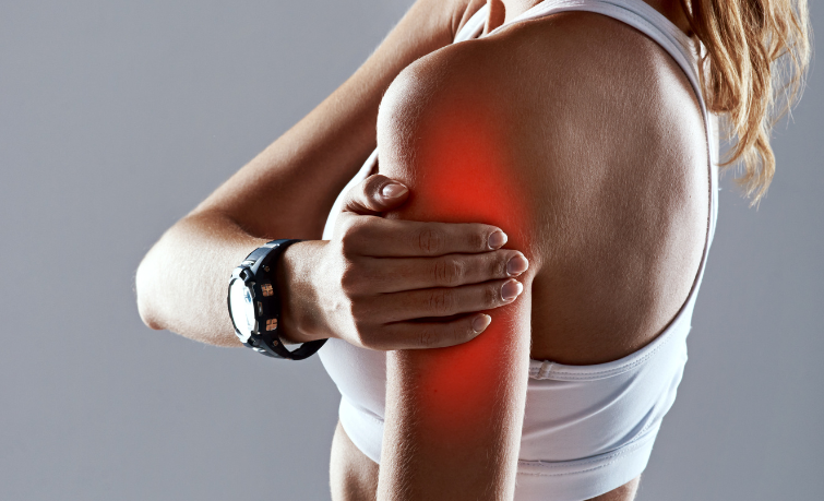
Typical symptoms of proximal biceps long head tendon pathology include pain in the anterior aspect of the joint, worsening with movement, which can also give a sensation of painful popping along the course of the biceps long head tendon over the humeral bone. All of these symptoms result in weakness of the tendon and muscle. In cases of complete tendon damage, there may be a deformity of the arm due to the biceps belly slipping down the humerus – Popeye sign.
Diagnosis
The diagnosis is made based on the history and clinical examination performed by a doctor, who uses specific tests to determine the pathology of the biceps long head tendon, its potential instability, and any accompanying injuries to the rotator cuff tendons (impingement with the subscapularis tendon) or labrum. Confirmation of the disease is obtained through ultrasound examination and magnetic resonance imaging (MRI).
Treatment of proximal biceps long head tendon pathology
The treatment of traumatic tendon damage, chronic overuse/inflammation, or accompanying symptomatic joint pathologies is surgical treatment.
The type of procedure performed depends on the type of damage. A typical procedure is arthroscopic surgery.
In the case of SLAP lesions, which involve damage to the labrum, including the attachment site of the biceps tendon, the treatment involves stabilizing (reattaching) the tendon along with the labrum to the glenoid surface using special anchors. This approach requires a good quality tendon. If this condition is not met, labrum stabilization is performed, and the tendon is detached at its attachment site in the upper part of the labrum. This procedure is called tenotomy and is also performed in cases of biceps tendon damage and instability accompanying rotator cuff tendon injuries. The result of tenotomy can be a biceps deformity resulting from the sliding of the muscle into the distal part of the arm and a slight weakening of its strength. For this reason, it is mainly used in older individuals with low demands for physical activity, overweight individuals, and those who are not bothered by any functional or aesthetic deficits.
An alternative procedure to tenotomy is tenodesis. This involves the reinsertion of the tendon onto the humerus bone.
The procedure can be performed at different levels using two methods:
- Arthroscopically, at the top of the groove or along its course on the humerus bone.
- Via an open approach, with an additional small incision in the groove or above/below the insertion of the pectoralis major tendon on the humerus bone.
In both cases, anchors are used to stabilize the tendon at the chosen location on the humerus bone.
Postoperative management includes temporary immobilization in a splint for 2-4 weeks. Rehabilitation begins in the first week after surgery and involves increasing the range of motion of the shoulder joint and exercising the elbow joint, gradually increasing muscle strength. The full rehabilitation process takes about 6 months.
It should be noted that the type and duration of immobilization, as well as the course and length of rehabilitation, depend on any additional procedures performed during the surgery.
In MIRAI, the shoulder area is taken care of by:
Bartosz Dominik – content development on the website
Michał Drwięga
You are welcome!