
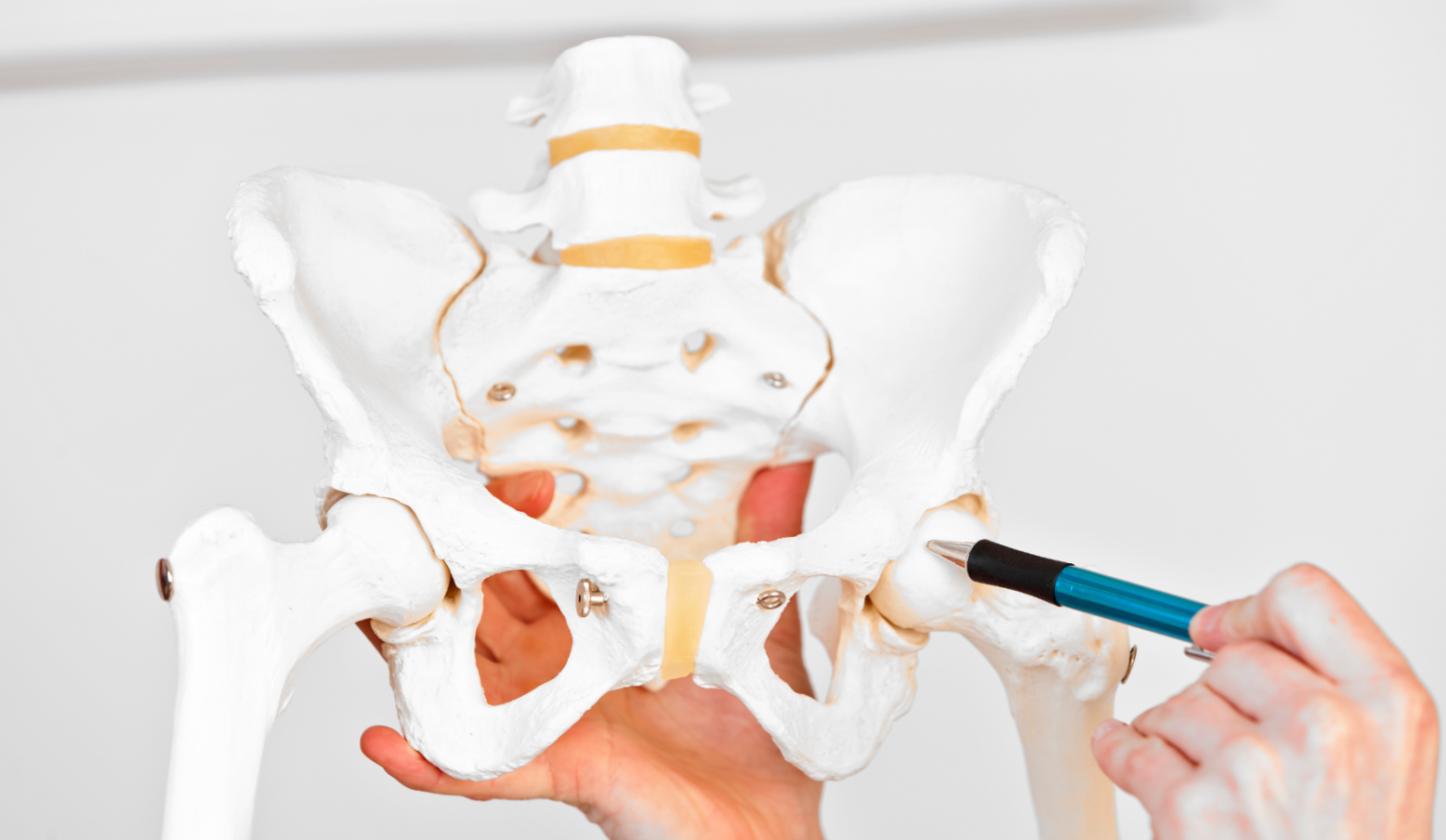
The hip joint is a ball-and-socket joint with multi-directional mobility. It allows movements such as flexion, extension, abduction, adduction, internal rotation, and external rotation. A total of 24 muscles are responsible for these movements. The hip joint consists of the femoral head and the acetabulum of the pelvic bone. The joint is surrounded by the joint capsule along with ligaments such as the iliofemoral ligament, pubofemoral ligament, ischiofemoral ligament, ligament of the head of the femur, and the circular layer. As you can see, there are many structures that can cause painful symptoms.
Hip Physiotherapy
Femoroacetabular Impingement (FAI)
FAI is an abnormal contact between the femoral head and acetabulum of the hip joint, caused by altered bone growth within the hip joint. FAI can be divided into 2 types:
- Cam-type impingement: Bone changes occur within the femoral head, leading to restricted rotation of the femoral head in the acetabulum of the hip joint.
- Pincer-type impingement: Bone changes occur in the acetabulum of the hip joint, leading to gradual damage to the articular cartilage.
- Mixed-type impingement: Occurs when bone alterations involve both the acetabulum of the hip joint and the femoral head.
Epidemiology: FAI develops due to overloads between the femoral neck, femoral head, and acetabulum, leading to excessive contact between these structures. It usually occurs in young individuals who are physically active. People who have had hip-related conditions in childhood, such as Perthes disease or hip dysplasia, are also more susceptible.
Symptoms: Hip joint pain, which may radiate to the thigh or spine, limited range of motion, audible “popping” or “clicking” in the hip joint (often when rising from a seated position).
Diagnosis: To diagnose the condition, a physiotherapist or orthopaedic specialist initially conducts a detailed patient interview, followed by a physical examination involving manual assessment and specific tests to identify the dysfunction. Imaging tests may also be performed to confirm the suspected diagnosis.
Treatment: In the early stages of the disease, conservative treatment is recommended, and in most cases, it yields positive results. Manual therapy, including hip joint mobilization and soft tissue relaxation, can provide relief to the patient. Properly tailored exercises are also essential to improve proprioception, muscle strength, and endurance.
When conservative treatment does not yield the desired results, intra-articular injections of hyaluronic acid or corticosteroids may be used to alleviate pain.
In advanced cases, surgical intervention may be necessary, during which the orthopaedic surgeon performs corrective bone surgery.
Degenerative Changes
Degenerative joint disease, also known as osteoarthritis, is a chronic non-inflammatory condition that involves an imbalance between the regeneration and degradation of joint cartilage and subchondral bone. Degenerative changes often occur with unknown causes (primary) or as a result of predisposing factors (secondary). Predisposing factors may include obesity, unequal limb length, lower limb axis abnormalities, childhood hip joint diseases, injuries, fractures, hip joint infections, avascular necrosis of bone, rheumatoid arthritis, Paget’s disease, or excessive strain from sports activities.
Epidemiology: The occurrence of degenerative changes increases with age. It most commonly affects older individuals, especially women. In Poland, degenerative changes in the hip joint account for 40% of all diagnosed degenerative changes.
Symptoms: Hip joint pain, which may radiate towards the knee, joint stiffness, limited range of motion, joint crepitus, and progressive loss of function, which can make daily activities challenging.
Diagnosis: Confirmation of the condition is based on imaging tests (X-rays), a detailed patient interview, and a physical examination using various tests. Blood tests may also be conducted to rule out infections and rheumatic diseases, joint fluid analysis, bone scintigraphy, and magnetic resonance imaging (MRI), especially in cases of suspected avascular necrosis.
Treatment for hip degeneration: Conservative treatment is usually the first-line approach. It primarily focuses on targeted elimination of functional impairments that may contribute to pain. These impairments include muscle weakness, muscle tightness, and impaired motor control in the trunk and lower limb. Additionally, physiotherapy procedures are utilized to complement the treatment and provide analgesic effects. Commonly used modalities include TENS (transcutaneous electrical nerve stimulation), high-energy laser therapy, and endogenous tissue heating therapy, which promotes tissue hyperemia and relaxation through deep tissue penetration. A proper diet that has anti-inflammatory properties and promotes weight normalization is also crucial. In cases of severe pain, joint injections of hyaluronic acid or autologous blood therapies may be recommended by an orthopedic specialist. When degenerative changes are advanced, hip joint arthroplasty (total hip replacement) may be performed.
Gluteus Medius Tendinopathy
Gluteus medius tendinopathy is a condition that arises from an imbalance between microtrauma and tendon repair. It can occur due to mechanical overload or metabolic disturbances. This leads to degenerative changes in the tendon, weakening its ability to transmit force and causing pain from the nerve endings within the tendon.
Epidemiology: Gluteus medius tendinopathy can affect individuals who are physically active as well as those with a sedentary lifestyle. It can develop from repetitive injuries over weeks or months, such as from altered running mechanics, or from a single mechanical stress event. An example of the latter would be an inactive person attempting a strenuous mountain hike. Gluteus medius tendinopathy is more common in women aged 40-60 years. The estimated prevalence in the general population is 10-25%.
Symptoms of gluteus medius tendinopathy: include pain in the greater trochanter region of the femur, which may radiate towards the knee joint or buttocks. The pain is often exacerbated when climbing stairs or crossing one leg over the other. Another symptom is difficulty standing on one leg, and swelling may occur in some cases. These symptoms significantly impact physical activity levels. Additionally, pain in the trochanteric region (lateral side of the hip) while lying on the side can lead to sleep disturbances.
Diagnosis: In addition to a detailed patient interview and physical examination, imaging tests can be helpful for diagnosis. Magnetic resonance imaging (MRI) is an effective method for assessing the structure of the gluteus medius and minimus tendons.
Physiotherapy plays a major role in restoring function, involving exercises to correct training errors, improve movement patterns, and gradually strengthen the weakened tendons. The exercises typically start with isometric work and collagen training, which involves performing a high number of repetitions without any load. Then, exercises with increasing resistance and slow movement tempo are introduced. Dynamic exercises are introduced at the end. In the case of gluteus medius tendinopathy, autologous blood injections may also be used. In severe cases with significant tendon changes, surgical intervention may be necessary.
Acetabular labral tear
The acetabular labrum is a cartilaginous structure that surrounds the socket of the hip joint, increasing its surface area, absorbing the forces on the hip joint, and providing stability.
Epidemiology: Acetabular labral tears most commonly occur in runners and soccer players due to cumulative microtrauma. They often affect individuals who have a history of multiple hip joint injuries or those with anatomical abnormalities that contribute to labral damage, such as hip dysplasia, labral hypertrophy, or femoroacetabular impingement.
Symptoms of acetabular labral tear: The most common symptom is pain in the groin area, which worsens during prolonged sitting, standing, walking, or physical activity. Patients may also experience a catching sensation deep within the joint, limping during walking, and limited range of motion. Occasionally, the Trendelenburg sign may be observed, which is the dropping of the pelvis on the opposite side of the affected hip during one-legged stance.
Diagnosis: Diagnosis is typically made through a combination of patient history, physical examination, and imaging studies such as magnetic resonance imaging (MRI) or magnetic resonance arthrography (MRA).
Treatment for acetabular labral tear: The treatment approach may vary depending on the severity of the tear and the patient’s symptoms. Conservative management options include rest, physical therapy, nonsteroidal anti-inflammatory drugs (NSAIDs), and activity modification. In cases where conservative treatment fails to alleviate symptoms, surgical intervention such as arthroscopy may be recommended to repair or remove the torn labrum.
You’re welcome!